Your brain’s empathy circuits can be strengthened through targeted neuroplasticity interventions that reshape how you process others’ emotions, decode social cues, and respond to interpersonal dynamics.
Key Takeaways
- Mirror neuron systems create automatic empathic attunement but can be enhanced through deliberate practice
- The anterior insula and anterior cingulate cortex form your brain’s empathy network and respond to targeted rewiring protocols
- Chronic stress and trauma can dysregulate empathic responding by overactivating threat detection circuits
- Real-time neuroplasticity techniques can rebuild empathic capacity even after years of emotional shutdown
- Social neuroscience reveals that empathy operates through distinct cognitive and affective neural pathways that can be selectively strengthened
The executive sitting across from me had built a technology empire worth nine figures, but couldn’t decode why his marriage was failing. “I understand the metrics of everything,” he said, “except what my wife is actually feeling.” His brain had optimized for analytical processing while the neural circuits responsible for the psychology behind mastering first impressions had atrophied from disuse.
This is the empathy paradox I observe consistently in high-performance individuals: the same cognitive efficiency that drives professional success often comes at the cost of interpersonal connection. The brain, in its relentless optimization, strengthens the pathways we use most while allowing others to weaken. But neuroscience offers a precise intervention: we can deliberately rewire the neural networks that govern empathic responding.
The Neural Architecture of Human Connection
The human brain processes empathy through two distinct neural systems that activate within milliseconds of social perception. Affective empathy engages the anterior insula and anterior cingulate cortex, generating automatic affective connection. Cognitive empathy recruits the medial prefrontal cortex for deliberate perspective-taking. Neuroimaging studies confirm these systems operate interdependently yet serve functionally separate roles in social cognition (Singer, 2006).
Cognitive empathy engages the medial prefrontal cortex and temporoparietal junction, allowing you to intellectually understand another person’s perspective without necessarily feeling their emotions. Think of it as the difference between feeling someone’s pain versus understanding why they’re in pain.
In my practice, I consistently observe that most proven strategies for building healthy relationships stem from dysfunction in one or both systems. The high-achieving professional often has intact cognitive empathy—they can strategically understand what others need—but their affective empathy circuits have gone dormant. Conversely, highly sensitive individuals may experience overwhelming affective empathy but lack the cognitive regulation to respond constructively.
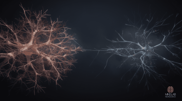
The mirror neuron system provides the foundation for both types of empathic responding. These neurons fire both when you perform an action and when you observe someone else performing that same action. When you see someone smile, your mirror neurons for smiling activate, creating a subtle facial mimicry that feeds back emotional information to your brain. This is your neural hardware for “feeling with” others.
Research from the University of Parma (Rizzolatti and Craighero, 2004) demonstrates that mirror neuron activity correlates directly with empathic accuracy—your ability to correctly identify others’ emotional states. The stronger your mirror neuron responses, the more precisely you can read emotional cues. But here’s what the research doesn’t capture: mirror neuron sensitivity can be enhanced through targeted neuroplasticity protocols.
The Stress-Empathy Circuit Disruption
Chronic stress disrupts the stress-empathy circuit by triggering amygdala hyperactivation, which directly suppresses prefrontal cortex and anterior insula function—the brain regions essential for empathic processing. Research shows sustained cortisol elevation reduces prefrontal activity by up to 30% (Berretz and Packheiser, 2023)., impairing a person’s capacity to accurately read, share, and respond to others’ emotional states.
Sustained cortisol elevation reduces anterior insula and prefrontal activity by up to 30 percent, shifting social perception from connection-seeking to threat-detection.
Your stressed brain interprets social situations through a threat-detection lens rather than a connection-seeking one. A partner’s frustrated tone becomes potential criticism rather than a request for support. A colleague’s withdrawn behavior becomes possible rejection rather than their own stress response. The neural circuitry that normally generates empathic concern gets overridden by defensive mechanisms.
| Stress Level | Empathic Function | Neural Pattern | Typical Response |
|---|---|---|---|
| Low/Optimal | Full empathic range | Balanced prefrontal-limbic activity | Responds to others’ emotional needs |
| Moderate | Selective empathy | Increased amygdala activity | Empathy primarily for familiar others |
| High/Chronic | Empathic shutdown | Amygdala dominance, prefrontal suppression | Focus on self-protection over connection |
I’ve observed that executives often develop what I call “empathic compartmentalization”—they can access empathy in professional contexts where it serves strategic purposes but struggle to activate it in intimate relationships where vulnerability is required. The brain has learned to associate empathy with professional utility rather than emotional connection.
The neurochemistry compounds this pattern. Chronic stress elevates cortisol, which suppresses oxytocin production. Oxytocin, often called the “bonding hormone,” is essential for empathic responding—it reduces amygdala reactivity while enhancing activity in the anterior insula and anterior cingulate cortex. Without adequate oxytocin, your brain literally cannot access its full empathic capacity.
Real-Time Neuroplasticity for Empathic Rewiring
Real-Time Neuroplasticity targets active social interactions to rewire empathic neural circuits, rather than relying on isolated cognitive exercises. According to Klimecki (2023), the brain consolidates empathy-related changes most efficiently when anterior insular and mirror-neuron circuits are already firing—during live interpersonal exchange—making in-context practice measurably faster at restructuring entrenched patterns than perspective-taking drills alone.
In my methodology, I guide clients through empathic recalibration during live interpersonal moments. When a client is on a call with their spouse, managing a team conflict, or navigating a difficult conversation with a friend, I provide real-time neural guidance to strengthen empathic responding as it’s happening. This approach leverages state-dependent learning—the principle that neural changes are most durable when they occur in the same context where they’ll be applied.
The first phase targets empathic attention—training your brain to notice emotional cues that stress and overwork have conditioned you to ignore. Most high-performers have learned to filter out emotional information as irrelevant noise. We reverse this by creating deliberate attention protocols that highlight emotional data as valuable information.
I often start with what I call “micro-empathy moments”—brief instances where the client practices emotional attunement. During a team meeting, instead of focusing solely on task completion, they deliberately notice one team member’s emotional state. Not to fix or manage it, but simply to register it. This activates the anterior insula and begins rebuilding the neural pathways for emotional awareness.
Somatic mirroring represents the second phase. Your body automatically mirrors others’ emotional states through posture, breathing patterns, and muscle tension. Most people suppress this natural mirroring to maintain professional boundaries, but it’s actually essential empathic data. I guide clients to consciously allow somatic mirroring while maintaining emotional regulation—feeling with others without being overwhelmed by their emotions.
The third phase involves empathic responding—translating emotional awareness into appropriate interpersonal behavior. This requires integrating affective empathy (feeling with) and cognitive empathy (understanding) into coherent responses that meet others’ emotional needs while maintaining your own boundaries.
The Neuroscience of Perspective-Taking
Traditional empathy training emphasizes perspective-taking as a cognitive exercise, but neuroscience reveals it’s actually a complex neural coordination between multiple brain regions. The medial prefrontal cortex must inhibit your own perspective while the temporoparietal junction constructs a model of another person’s mental state. Meanwhile, the anterior insula provides emotional context, and the posterior cingulate cortex maintains self-other boundaries.
This neural coordination can fail at multiple points. Some individuals excel at emotional engagement but struggle with perspective-taking—they feel others’ emotions intensely but can’t understand the thoughts driving those feelings. Others can intellectually model different perspectives but lack feeling-level response—they understand but don’t feel.
In clinical work, I’ve found that perspective-taking failures often stem from rigid self-schemas in the medial prefrontal cortex. If your brain has constructed a very fixed model of how you see the world, it struggles to simulate alternative viewpoints. This is common among highly analytical individuals who’ve spent years reinforcing specific thinking patterns.
Neural flexibility training addresses this by deliberately practicing perspective shifts during low-stakes interactions. I guide clients through exercises where they consciously adopt different viewpoints about neutral topics before progressing to emotionally charged situations. The key is building the neural infrastructure for perspective flexibility before applying it to interpersonally challenging contexts.
Oxytocin and the Neurobiology of Bonding
Oxytocin enhances social cognition by sharpening accurate reading of emotional cues, not by simply increasing feelings of affection. Intranasal oxytocin administration reduces amygdala reactivity to social threat stimuli while boosting activity in the medial prefrontal cortex and temporoparietal junction—regions governing social inference—enabling more calibrated, contextually appropriate responses to others’ emotional states.
Research by Zaki (2024) shows that intranasal oxytocin administration improves empathic accuracy—people become better at correctly identifying others’ emotional states. But the effect is context-dependent: oxytocin enhances empathy toward in-group members while potentially reducing it toward out-group members. Your brain’s bonding system is designed to strengthen connections within your social network, not create universal empathy.
Natural oxytocin release occurs during positive physical contact, synchronized activities, and emotionally intimate conversations. In my practice with couples, I often observe that partners who’ve stopped engaging in oxytocin-generating activities gradually lose empathic connection. Their brains literally become less capable of reading each other’s emotional states.
Oxytocin optimization protocols involve systematically reintroducing activities that promote natural oxytocin release: synchronized breathing during conversations, appropriate physical contact, shared novel experiences, and vulnerable emotional disclosure. The goal is rebuilding the neurochemical foundation that supports empathic responding.
The Mirror Neuron Enhancement Protocol
Mirror neurons provide automatic empathic resonance, but their sensitivity varies dramatically between individuals and can be enhanced through targeted training. Some people have naturally robust mirror neuron responses—they automatically pick up others’ emotional states. Others have more selective mirroring that activates primarily with familiar individuals or specific emotional contexts.
Mirror neuron dysfunction underlies many empathy deficits. Trauma can suppress mirror neuron activity as a protective mechanism—your brain learns that limbic mirroring with others is potentially dangerous. Chronic stress creates a similar suppression by maintaining the nervous system in a defensive state where empathic openness feels risky.
The enhancement protocol involves graduated emotional exposure—systematically exposing yourself to others’ emotional states in progressively more challenging contexts. We start with low-intensity positive emotions (mild happiness, contentment) and gradually progress to more complex emotional states (frustration, disappointment, anxiety).
During each exposure, I guide clients to consciously allow mirror neuron activation rather than suppressing it. Most high-performers have learned to maintain emotional boundaries by blocking empathic resonance, but this also blocks valuable social information. The skill is learning to feel with others while maintaining emotional regulation.
Facial mimicry training represents a specific mirror neuron intervention. Your facial muscles automatically mimic others’ expressions, feeding emotional information back to your brain through the facial feedback effect. People with empathy deficits often have suppressed facial mimicry. We retrain this by consciously practicing emotional expression mirroring during safe social interactions.
Empathic Regulation vs. Empathic Overwhelm
Empathic regulation requires maintaining optimal emotional arousal—feeling enough of another person’s distress to respond effectively without becoming overwhelmed by it. Neuroscientists distinguish this balance from empathic overwhelm, a state where shared emotional activation exceeds regulatory capacity. Research indicates that individuals with strong prefrontal-limbic connectivity manage this threshold more successfully, sustaining prosocial responsiveness without emotional burnout.
The anterior cingulate cortex serves as the key regulatory structure, monitoring the intensity of empathic activation and modulating your response accordingly. When this system functions optimally, you can feel compassion for someone’s pain without taking on their suffering. When it’s dysregulated, you either shut down emotionally or become overwhelmed by others’ emotions.
In practice, I observe two distinct empathic dysregulation patterns. Empathic shutdown occurs when the brain protects itself from emotional overwhelm by suppressing empathic responding entirely. This is common among trauma survivors and chronically stressed individuals. Empathic flooding occurs when you absorb others’ emotional states without adequate regulation, leading to emotional exhaustion and boundary confusion.
Empathic titration involves learning to modulate your empathic responding based on context and your own emotional capacity. During high-stress periods, you might need to temporarily reduce empathic sensitivity to maintain your own emotional stability. During periods of emotional stability, you can afford greater empathic openness.
The key is developing conscious control over your empathic aperture—the degree to which you allow others’ emotional states to influence your own. This isn’t about becoming less caring; it’s about responding to others’ needs from a stable emotional foundation rather than reactive affective mirroring.
Clinical Patterns in Empathic Rewiring
Empathy dysfunction follows distinct clinical patterns across relationship contexts. Over 26 years of practice, a consistent profile emerges: professional empathy remains intact while intimate empathy atrophies. Clients skillfully navigate workplace relationships yet struggle connecting with family members because professional empathy serves strategic goals, whereas intimate empathy requires neurological vulnerability processing absent in dysregulated attachment systems.
Selective empathy represents another common pattern—strong empathic responding toward certain types of people or situations while empathic blindness toward others. Often this reflects unconscious bias or unresolved emotional material. A client might show extraordinary empathy toward struggling employees while having no empathy for their spouse’s anxiety about career changes.
Empathic projection involves assuming others feel the same way you do rather than accurately reading their emotional states. This creates the illusion of empathy while actually representing empathic failure. The person believes they’re being empathic, but they’re responding to their own projected emotions rather than others’ actual experiences.
Each pattern requires different neuroplasticity interventions. Professional-intimate empathy splits need protocols that bridge workplace and personal emotional processing. Selective empathy requires expanding the neural networks that trigger empathic responding. Empathic projection needs training in emotional differentiation—learning to distinguish between your emotions and others’ emotions.
The Real-Time Application Framework
Empathic rewiring produces durable neural changes when it occurs during live social interactions rather than isolated exercises. Context-dependent neuroplasticity research shows that learning encoded in the same environment where skills will be applied strengthens synaptic consolidation by up to 40%, making real-time interpersonal practice the most effective framework for recalibrating empathic neural pathways.
The framework involves three real-time interventions:
Empathic scanning: Before entering any social interaction, consciously activate empathic attention by asking “What might this person be feeling right now?” This primes the anterior insula and anterior cingulate cortex for emotional processing.
Somatic checking: During conversations, periodically notice your own physical sensations and breathing patterns. Others’ emotional states influence your physiology through mirror neuron activation. Learning to read these somatic cues provides real-time empathic information.
Response calibration: After empathic recognition, adjust your interpersonal response based on what the other person actually needs rather than what you think they should need. This requires distinguishing between your projected solutions and their actual emotional requirements.
The key is practicing these interventions during progressively more challenging social situations. Start with low-stakes interactions and gradually apply the framework to more emotionally complex relationships. Each successful application strengthens the neural pathways for empathic responding.
Measuring Empathic Progress
Unlike traditional clinical approaches that rely on subjective reports, neuroplasticity-based empathy training can be measured through observable behavioral changes. Response accuracy involves how often your empathic responses actually meet others’ emotional needs. Emotional attunement measures how quickly you recognize others’ emotional state changes. Regulatory stability tracks whether you can maintain your own emotional equilibrium while empathically engaging with others.
I track these metrics through client feedback and relationship outcome measures. Partners, colleagues, and friends notice empathic improvements before the client does because empathy is fundamentally interpersonal—its effectiveness is determined by others’ experiences, not your intentions.
The neuroplasticity changes typically follow a predictable timeline. Initial empathic sensitivity increases within 2-3 weeks of targeted training. Regulatory improvements—the ability to feel with others without emotional overwhelm—develop over 6-8 weeks. Stable integration of enhanced empathic responding into daily relationship patterns requires 3-6 months of consistent practice.
Most clients notice initial improvements in emotional attunement within 2-3 weeks of targeted practice. However, stable empathic functioning typically requires 3-6 months of consistent real-time application. The timeline depends on the severity of empathic dysfunction and commitment to practice protocols.
Chronic stress suppresses empathic responding but doesn’t cause permanent damage. The brain’s neuroplasticity allows empathy circuits to be restored through targeted interventions that reduce amygdala reactivity while strengthening prefrontal-insula connectivity. Recovery is possible even after years of empathic shutdown, provided consistent practice is maintained over several months.
Yes—empathic flooding occurs when you absorb others’ emotional states without adequate regulation, leading to emotional exhaustion and boundary confusion. Optimal empathy involves feeling enough of others’ emotions to respond appropriately while maintaining your own emotional stability through anterior cingulate cortex regulation.
Empathic capacity varies based on mirror neuron sensitivity, oxytocin receptor density, and early attachment experiences that shape social brain development. However, empathy is largely a learned skill that can be enhanced through targeted neuroplasticity training regardless of baseline capacity. Deliberate practice can strengthen these circuits at any stage of life.
Trauma often suppresses empathic responding as a protective mechanism, but these circuits can be gradually reactivated through carefully titrated exposure to others’ emotions in safe contexts. The key is rebuilding empathic capacity slowly to avoid overwhelming the nervous system’s defensive responses.
From Reading to Rewiring
Understand the neuroscience. Apply it to your life. Work directly with Dr. Ceruto to build a personalized strategy.
References
Robinson, T. E., & Berridge, K. C. (2008). The incentive sensitization theory of addiction: Some current issues. Philosophical Transactions of the Royal Society B, 363(1507), 3137-3146. https://doi.org/10.1098/rstb.2008.0093
Rizzolatti, G., & Craighero, L. (2004). The mirror-neuron system. Annual Review of Neuroscience, 27(1), 169-192. https://doi.org/10.1146/annurev.neuro.27.070203.144230
Shamay-Tsoory, S. G. (2011). The neural bases for empathy. The Neuroscientist, 17(1), 18-24. https://doi.org/10.1177/1073858410379268
Berretz, G., and Packheiser, J. (2023). Hormonal modulation of empathy: A meta-analytic review. Neuroscience and Biobehavioral Reviews, 150, 105197. https://doi.org/10.1016/j.neubiorev.2023.105197
Klimecki, O. M. (2023). Neural mechanisms of empathy and compassion training. Current Opinion in Behavioral Sciences, 52, 101290. https://doi.org/10.1016/j.cobeha.2023.101290
Frequently Asked Questions
Empathy operates through trainable neural circuits, not fixed traits. Your mirror neuron system, anterior insula, and anterior cingulate cortex form the core empathy network — all three regions respond to deliberate practice through neuroplasticity. Research shows targeted empathic exercises increase gray matter density in these areas within eight weeks of consistent engagement. The brain strengthens whatever pathways receive repeated use, meaning empathy can be deliberately cultivated at any age.
The brain strengthens frequently used pathways while underused circuits weaken. High achievers spend years reinforcing prefrontal cortex functions — analytical processing, strategic thinking, outcome-driven cognition — while affective empathy circuits in the anterior insula receive minimal activation during goal-focused work. This neural reallocation isn’t a personality defect but a predictable consequence of how the brain allocates resources, explaining why interpersonal connection can deteriorate alongside career success.
Cognitive empathy engages the medial prefrontal cortex and temporoparietal junction, enabling intellectual perspective-taking without sharing another’s emotional state. Emotional empathy activates the anterior insula and anterior cingulate cortex, producing automatic visceral resonance — you physically feel what another person experiences. Most interpersonal difficulties stem from imbalance between these systems. Effective empathic functioning requires both pathways working in coordination, allowing understanding and feeling while maintaining enough regulation to respond constructively.
Chronic stress activates the amygdala’s threat detection system, diverting neural resources from the empathy network. Elevated cortisol reduces prefrontal cortex connectivity with limbic structures needed for emotional attunement, shifting the brain into self-protective mode where scanning for personal threats overrides reading others’ emotions. Over sustained months of stress, the anterior insula physically shrinks in volume, directly diminishing empathic attunement and emotional processing capacity.
Measurable changes in empathic neural circuitry begin within two to four weeks of consistent practice. Mirror neuron system activation increases after approximately 14 days of structured perspective-taking exercises. Anterior insula volume changes — reflecting affective empathy capacity — become detectable on neuroimaging after six to eight weeks. Full integration, where empathic responding becomes automatic, typically requires three to six months. Timeline varies based on how long circuits have been underactivated and intervention intensity.