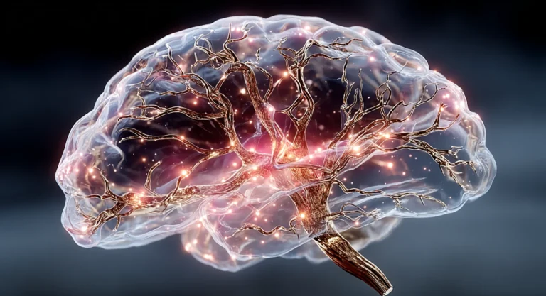
Neuroinflammation and Cognitive Decline: What IL-6, TNF-alpha, and CRP Reveal About Your Brain’s Inflammatory Load
Neuroinflammation symptoms in the brain rarely announce themselves as inflammation. They arrive as slower word retrieval, reading the same paragraph twice, and decision fatigue disproportionate to workload. The inflammatory markers driving these shifts — interleukin-6, tumor necrosis factor alpha, and high-sensitivity C-reactive protein — each predict cognition with very different accuracy.
Key Takeaways
- IL-6 is the strongest midlife cognitive predictor. It partially mediates age-related processing-speed decline where TNF-alpha and CRP do not, and it crosses the blood-brain barrier at low concentrations.
- High-sensitivity C-reactive protein is the weakest cognitive predictor despite being the most commonly ordered test. It reflects systemic inflammation but does not independently track cognitive trajectory.
- Brain inflammation presents as cognitive slippage, not classic inflammation signs. Redness, heat, and swelling describe peripheral tissue; neuroinflammation shows up as retrieval delay, working-memory compression, and executive fatigue.
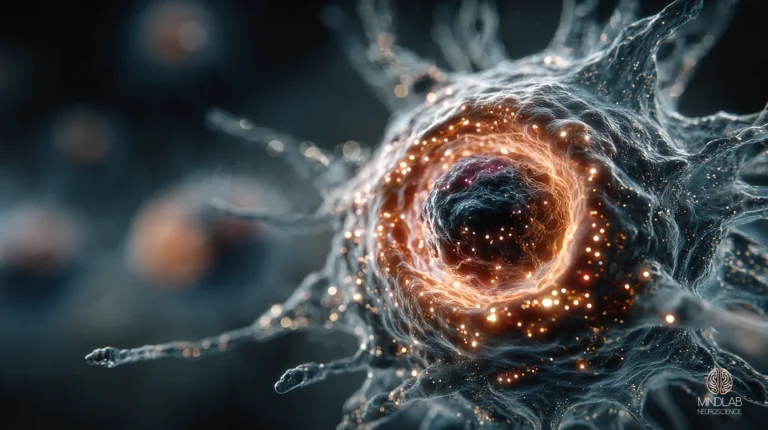
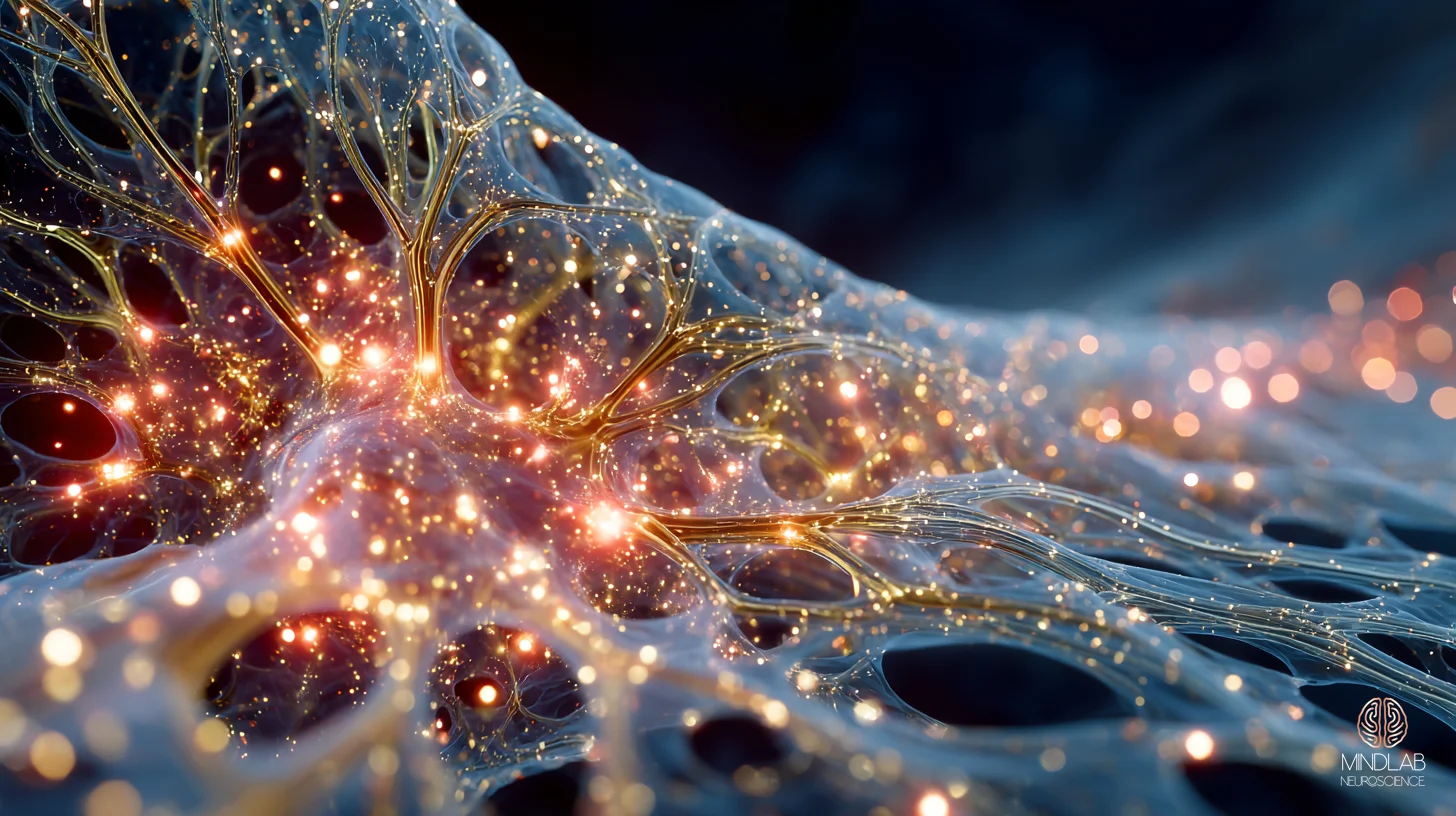
- Microglial activation is the cellular signature. Activated microglia drive chronic cytokine output inside the brain even when systemic markers look unremarkable.
- Peripheral inflammation converts into central inflammation through blood-brain barrier disruption. Sleep quality, dietary inflammatory load, and visceral fat quietly feed the pathway without anyone calling it “brain inflammation.”

How Do You Know If You Have Brain Inflammation?
Brain inflammation rarely produces the classic symptoms people associate with inflammation elsewhere in the body. It shows up as sustained cognitive slippage across domains — verbal retrieval delays, slower reading comprehension, decision fatigue disproportionate to workload, and a working-memory ceiling that keeps dropping. Quantitative markers then confirm what the client reports.
In my practice, I consistently observe this pattern in professionals who have been operating at their usual standard and then notice a year of quiet slippage. They come in believing they need better sleep, more exercise, or sharper focus. The lab panel tells a different story. A 32-year-old attorney came to me after transactions that used to take her half a day started taking a full day. Her fasting IL-6 was elevated at 3.4 pg/mL. Her high-sensitivity CRP, drawn twice in six months, read inside range both times. The cognitive signature preceded any marker her internist was watching.
Published data agrees with what the clinic shows. A 2018 study (Lin et al., Frontiers in Aging Neuroscience) found that IL-6 partially mediated age-related processing-speed decline even in generally healthy adults, while TNF-alpha and CRP showed no comparable mediation effect in the same cohort. IL-6 was doing work that CRP was not.
This is why “Is my brain inflamed?” is a poor question. The better question is which marker best predicts what you are feeling, and whether the pattern is central, peripheral, or converting from one to the other.
What Tests Show Inflammation in the Brain?
No peripheral blood test shows inflammation inside the brain directly. Serum IL-6 is the strongest indirect predictor, outperforming TNF-alpha and high-sensitivity CRP in mediation analyses of cognitive decline. Each marker measures a different layer of the inflammatory signal. The most commonly ordered test — hs-CRP — is the weakest standalone cognitive predictor.
The standard protocol recommends ordering hs-CRP. In 26 years I’ve found hs-CRP tells you the body is inflamed; it rarely tells you which markers are driving cognition. IL-6 is a pro-inflammatory cytokine produced by immune cells, adipose tissue, and stressed muscle. It is small enough to cross the blood-brain barrier through saturable transport. TNF-alpha tracks cross-sectional cognitive deficits but lacks the predictive specificity for trajectory. High-sensitivity CRP is a downstream liver-produced acute-phase reactant and sits at the end of the pathway, not the beginning.
A 2022 meta-analysis (Custodero et al., GeroScience) synthesized thirteen cross-sectional and seven prospective studies examining IL-6, CRP, and TNF-alpha in vascular cognitive impairment. Blood IL-6 discriminated vascular dementia from Alzheimer’s disease with a standardized mean difference of 0.40 (95% CI 0.18–0.62) and predicted incident vascular dementia with a relative risk of 1.28 (95% CI 1.03–1.59). CRP and TNF-alpha lacked comparable discriminant validity in the same pooled analysis.
“The standard protocol recommends hs-CRP. In 26 years I’ve found hs-CRP tells you the body is inflamed — it rarely tells you which markers are actually driving cognition.”
The practical implication matters. A client with a normal CRP and an elevated IL-6 has a different cognitive trajectory, a different set of levers, and a different working-memory ceiling than a client with the reverse pattern. The panel is not interchangeable, and ordering the wrong one first is how a year of cognitive slippage gets labeled “anxiety” or “workload.”
Does High Inflammation Cause Brain Fog?
Yes — when the inflammatory signal crosses the blood-brain barrier. Brain fog is the felt experience of cytokine activity inside the central nervous system. Elevated IL-1β, TNF-alpha, and IL-6 disrupt blood-brain barrier integrity, allowing peripheral inflammation to register centrally as cognitive impairment. The process is mechanistic, not metaphorical.
Brain fog is not a vague state. It is a specific cognitive phenotype: delayed retrieval, reduced filtering of irrelevant input, and a thinner working-memory margin under time pressure. When I work with midlife executive clients, the pattern repeats itself almost identically. A 52-year-old head of strategy at a midsize firm came in running fine by most observable metrics but quietly losing access to his own analytical precision. He could still hold three competing scenarios in working memory; he could no longer hold four the way he used to. His IL-6 was 4.6 pg/mL. His hs-CRP was inside range.
A 2022 review (Yang et al., Frontiers in Molecular Neuroscience) documented how IL-1β, TNF-alpha, and IL-6 destabilize tight-junction proteins in the blood-brain barrier, permitting peripheral cytokines to register centrally and produce the cognitive signature clients describe as fog. Complementary human data (Skoczek-Rubińska et al., 2021) found that women already showing cognitive impairment had IL-6 concentrations 64% higher than cognitively normal peers (4.1 versus 2.5 pg/mL, p = 0.004) — the marker was elevated even when the subjective experience felt like ordinary fatigue.
What the research does not fully capture is that not every brain fog is neuroinflammatory. Sleep restriction produces similar subjective experience without cytokine elevation. Confirmation requires the lab pattern plus the cognitive phenotype plus the trajectory — three data layers, not one.
How Do You Reverse Neuroinflammation?
Neuroinflammation reverses through targeted action on the peripheral-to-central pathway — sleep quality, dietary inflammatory load, visceral adiposity, and BDNF-supporting input. The inflammatory brain cannot efficiently rewire because IL-1β suppresses BDNF-dependent synaptic plasticity. Restoring cognitive function requires addressing the inflammatory load first, then supporting the underlying neural architecture.
Four levers carry most of the clinical change I observe: sleep architecture, dietary inflammatory load, visceral fat, and BDNF-supporting input (movement, omega-3 fatty acids, and sustained cognitive demand at the edge of ability). Sleep sits first because the evidence is strongest. Prolonged sleep deficiency produces chronic low-grade systemic inflammation with elevated IL-6 and TNF-alpha, and the effect appears at sleep durations clients consider normal (Besedovsky et al., 2019). Diet sits second. Each one-point increase in the Dietary Inflammatory Index corresponded to 1.55-times greater odds of cognitive impairment in postmenopausal women (Skoczek-Rubińska et al., 2021) — the anti-inflammatory gradient is graded, not binary.
The highest-leverage case I see most often is not the C-suite executive. A 42-year-old came to me managing a mother with early dementia, a 14-year-old son in a hard school year, and a board seat she had not yet resigned from. She presented with executive fatigue, which her internist had screened six weeks earlier with a normal hs-CRP. Her IL-6 was 4.2 pg/mL. Her sleep actigraphy showed 5.2 hours average with 24 minutes of wake after sleep onset. The pathway was obvious once named: chronic sleep debt, elevated cortisol, disrupted cytokine rhythm, IL-6 signature. The inflammatory load was not from corporate decision volume. It was from the structural weight of managing three generations simultaneously — and it responded to sleep restoration within eight weeks.
The peripheral inflammatory signature responds to intervention. In my practice, Real-Time Neuroplasticity™ engagement begins only after the inflammatory load has been addressed at the systemic level, because IL-1β and its downstream cytokines suppress BDNF-dependent long-term potentiation — the cellular mechanism by which the brain consolidates new wiring. An inflamed brain cannot efficiently rewire. The order of operations matters.
What Are the 5 Classic Signs of Inflammation?
The five classical signs — redness, heat, swelling, pain, and loss of function — describe peripheral tissue inflammation from the Roman encyclopedist Celsus, later expanded by Galen. Neuroinflammation does not present any of them visibly. The brain has no visible surface, no skin, and its ‘loss of function’ manifests as cognition rather than as mobility or pain.
The standard answer to “what are the 5 classic signs” is useful for a cut on your arm. It is misleading for what is happening inside your skull. The cellular signature of neuroinflammation (Skaper et al., 2018; Heneka et al., 2012) involves microglial activation, astrocyte reactivity, NLRP3 inflammasome engagement, and cytokine output — none of which produce redness or heat in any observable sense. What the brain gives you instead is the cognitive phenotype described in the preceding sections: retrieval delay, working-memory compression, filtering loss, decision fatigue.
When a client asks whether they have brain inflammation, I translate the question. We are not looking for the five classical signs. We are looking for the marker stratification (IL-6 leading), the cognitive phenotype (retrieval, filtering, working memory), and the trajectory (stable, worsening, or responsive to intervention). That is the map that changes outcomes.
References
Besedovsky, L., Lange, T., & Haack, M. (2019). The Sleep-Immune Crosstalk in Health and Disease. Physiological Reviews. https://doi.org/10.1152/physrev.00010.2018
Heneka, M. T., Kummer, M. P., Stutz, A., Delekate, A., & Schwartz, S. (2012). NLRP3 is activated in Alzheimer’s disease and contributes to pathology in APP/PS1 mice. Nature. https://doi.org/10.1038/nature11729
Skaper, S. D., Facci, L., Zusso, M., & Giusti, P. (2018). An Inflammation-Centric View of Neurological Disease: Beyond the Neuron. Frontiers in Cellular Neuroscience. https://doi.org/10.3389/fncel.2018.00072
Skoczek-Rubińska, A., Muzsik, A., Chmurzyńska, A., Jamka, M., & Walkowiak, J. (2021). Inflammatory Potential of Diet Is Associated with Biomarkers Levels of Inflammation and Cognitive Function among Postmenopausal Women. Nutrients. https://doi.org/10.3390/nu13072323
What the First Conversation Looks Like
When someone brings me a labs panel with a normal CRP and a clear sense that something has shifted in their cognition, the first conversation is about mapping, not ordering more tests. I want to understand the timeline of the slippage, the trajectory of the inflammatory markers in context, and where the peripheral signal is entering the central system.
We’ll look at sleep architecture, dietary inflammatory load, metabolic inflammation markers, and the specific cognitive phenotype — working-memory ceiling, retrieval latency, filtering capacity — to build the picture. From that picture we identify where a small, targeted shift changes the most downstream. Most people leave that first conversation knowing something specific about their own inflammatory signature they had not named before.
Frequently Asked Questions
Can you have neuroinflammation with a normal CRP?
Yes, commonly. High-sensitivity CRP is a systemic inflammation marker — not a brain-specific one — and in mediation analyses of age-related cognitive decline, CRP contributes little or nothing independent of IL-6. A normal CRP does not rule out elevated IL-6 or TNF-alpha, and it cannot rule out central microglial activation, which no routine serum panel measures directly. The pattern across markers matters more than any single reading, particularly at midlife when quiet cognitive slippage is most detectable and most responsive to intervention.
Is brain fog always caused by neuroinflammation?
No. Brain fog is a cognitive phenotype — delayed retrieval, filtering loss, working-memory compression — that has several mechanistic drivers. Sleep restriction alone can produce it without elevated cytokines. Hormonal transitions, thyroid dysfunction, chronic dehydration, and iron deficiency produce overlapping signatures. Confirming a neuroinflammatory cause requires the marker pattern (IL-6 elevation, BBB-disruption signals) plus the cognitive phenotype plus the trajectory — three data layers together, not a single lab reading in isolation.
How does IL-6 get into the brain?
IL-6 crosses the blood-brain barrier through saturable transport at low concentrations and contributes to BBB permeability at higher concentrations, compromising the tight-junction proteins that normally gate the central nervous system. Chronic low-grade elevation — the midlife pattern — is particularly problematic because it operates continuously rather than acutely. The peripheral-to-central pathway is most active where the BBB is thinnest and where peripheral inflammatory input (visceral fat, sleep debt, diet) sustains the signal over months and years rather than hours.
Do anti-inflammatory diets actually reduce brain inflammation?
The evidence supports graded improvement, not binary reversal. Each one-point increase in the Dietary Inflammatory Index corresponds to about 1.55-times greater odds of cognitive impairment in studied populations, which means each one-point decrease moves the needle in the opposite direction. Mediterranean-pattern eating, omega-3 adequacy, and visceral-fat reduction each move IL-6 measurably over weeks to months. The dietary lever is real, but it works alongside sleep and movement — it rarely reverses a sustained inflammatory signature in isolation.
How long does it take to reduce neuroinflammation markers?
Serum IL-6 responds measurably to sleep restoration within four to eight weeks when the underlying sleep architecture is addressed, not merely duration. Dietary inflammatory load shifts over a similar window. Cognitive phenotype improvement typically lags the marker change by two to four weeks because neural consolidation requires the BDNF-dependent plasticity window that the inflammatory load was suppressing. The sequence is marker first, then cognitive recovery — and the gap between them is diagnostically informative rather than discouraging.