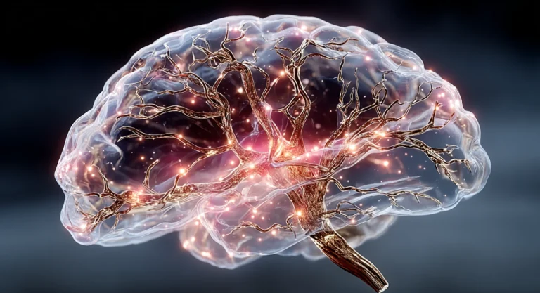
Mitochondrial Dysfunction in Neurons: How Cellular Energy Failure Drives Cognitive Decline
Key Takeaways
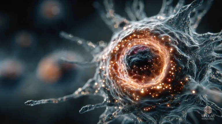
- Mitochondrial dysfunction in neurons is an electron transport chain failure, Complex I and Complex III lose catalytic integrity, ATP output falls, and reactive oxygen species leak into the cytosol where they damage membranes, proteins, and DNA.
- The unique cost is not just energy, it is neural stem cell depletion. Damaged mitochondria shift hippocampal stem cells out of self-renewal into premature differentiation, eliminating adult neurogenesis and the substrate of new learning.
- The signature is measurable in the periphery. PBMC mitochondrial panels, ATP production rate, basal respiration, proton leak, correlate with central bioenergetic status and are the earliest accessible readout of the dysfunction pattern.
- Repair is not supplementation. The PGC-1alpha / SIRT1 biogenesis axis rebuilds mitochondrial mass, and the axis activates under two conditions: sustained exercise above the lactate threshold, and caloric-restriction signaling.
- At MindLAB Neuroscience, I consistently find that adults arriving with “brain fog” that nootropic stacks and sleep protocols do not move are running the mitochondrial-dysfunction pattern, and that the pattern responds to biomarker-guided intervention, not symptom management.
Mitochondrial dysfunction in the brain is a progressive failure of neuronal ATP production, driven by electron transport chain Complex I and III impairment, that depletes the adult neural stem cell pool, collapses hippocampal neurogenesis, and produces a cognitive signature measurable in peripheral blood mononuclear cells through proton leak and ATP-production panels. The damage is not diffuse fatigue. It is architectural.


What Are the Symptoms of Damaged Mitochondria?
The cognitive signature of damaged mitochondria is specific, not generic. It presents as a progressive drop in processing speed, short-term working-memory failures under sustained load, and post-exertional cognitive crashes that outlast the physical fatigue by hours. The pattern does not resolve with a weekend of sleep, and it does not respond to stimulants. This article is part of the broader hub on the neuroscience of brain health and optimization, which connects the brain’s energy supply to the structures that depend on it.
The Afternoon Collapse That Rest Does Not Fix
A 31-year-old program manager at a venture-backed health-tech company came in convinced her 3 p.m. cognitive collapse was a sleep-debt problem. She had moved from seven to nine hours of sleep over eight weeks. She had cut evening alcohol. She had rotated through two nootropic stacks. Her morning window remained sharp; her 3 p.m. block felt 30 to 40 percent offline and no longer recovered before evening. The input pattern she was adding did not match the mechanism failing.
What she was describing is the canonical bioenergetic signature. When neuronal mitochondria fail, hippocampal and prefrontal circuits lose the sustained ATP supply that working memory requires to hold and manipulate information across seconds and minutes. The proximate mechanism is not fatigue in the muscular sense, it is the collapse of the oxidative phosphorylation flux that keeps synaptic vesicle cycling and ion-pump restoration running.
Why the Post-Exertional Crash Is Diagnostic
Post-exertional cognitive crash is the signal that distinguishes mitochondrial dysfunction from generic burnout. Exercise, cognitive effort, or even a demanding conversation increases neuronal ATP demand. A healthy system meets the demand and recovers within minutes. A mitochondria-compromised system meets the demand by drawing down already-depleted reserves, and the recovery window stretches to hours or longer. The literature on myalgic encephalomyelitis and post-acute sequelae of SARS-CoV-2 has documented the same pattern, shared energy-metabolism and redox abnormalities underlie both conditions and overlap substantially with the cognitive-decline population (Komaroff & Lipkin, 2023, see References).
The nuance: the symptom signature is not itself sufficient to conclude mitochondrial dysfunction. It is sufficient to warrant a bioenergetic workup. In my work at MindLAB, the next step is always a PBMC mitochondrial panel, basal respiration, ATP-linked respiration, proton leak, and spare respiratory capacity, before any intervention is designed.
How Does Mitochondrial Dysfunction Deplete the Neural Stem Cell Pool?
Mitochondrial dysfunction depletes the adult neural stem cell pool by disrupting the fate-decision machinery that keeps stem cells in self-renewal. When mitochondrial function fails, stem cells exit the quiescent state prematurely, differentiate into committed progenitors, and the pool progressively empties, eliminating adult hippocampal neurogenesis and the substrate of new learning.
The 2017 Finding That Changed the Frame
The mechanism was resolved in a 2017 paper in Human Molecular Genetics by Mireille Khacho and colleagues, who used conditional deletion of AIF, the mitochondrial oxidoreductase required for Complex I stability, to produce a genetic model of Complex-I deficiency in adult neural stem cells. The result was not diffuse neuronal death. It was a precise fate-decision failure: stem cells lost self-renewal capacity, exited the cell cycle prematurely, committed to a neuronal lineage too early, and the adult neural stem cell pool was eliminated within weeks. Cognitive performance on hippocampus-dependent tasks collapsed in parallel.
This is the reframe that most commodity coverage misses. Mitochondrial dysfunction does not simply make existing neurons sluggish. It empties the reservoir of cells that the hippocampus uses to encode new episodic memory, and it does so before the mature neuronal pool shows detectable damage.
“Damaged mitochondria do not just tire the brain. They empty the reservoir, the stem cell pool your hippocampus uses every day to encode new memory, and they do it before any mature neuron shows visible damage.”
A Non-Corporate Case, a Shared Mechanism
A woman managing a blended household, an aging parent’s care coordination, and a volunteer board role described a working-memory “grayness” that new sleep and calorie changes could not shift. She was not a corporate executive. She did not frame herself as a high-performer. What she described was the exact hippocampal-encoding deficit the NSC-depletion model predicts, new information refused to settle, and recent conversations felt porous by the next morning.
The mechanism does not care about job title. The hippocampus needs an adult neural stem cell pool to keep encoding new material, and that pool needs mitochondrial integrity to stay in self-renewal. Remove either and the encoding fails. Raising BDNF naturally runs a parallel architecture, BDNF-TrkB signaling supports plasticity while PGC-1alpha / SIRT1 supports the energetic substrate; both must be intact for new learning to stick.
How Do You Repair Depleted Mitochondria?
Depleted mitochondria are repaired primarily through mitochondrial biogenesis, the construction of new mitochondria via the PGC-1alpha / SIRT1 transcriptional axis, combined with mitophagy, the selective autophagy of damaged mitochondria. Both processes run continuously, and the intervention target is the upstream signaling that activates them.
The PGC-1alpha / SIRT1 Axis Is the Lever
PGC-1alpha, peroxisome proliferator-activated receptor gamma coactivator 1-alpha, is the master transcriptional coactivator for mitochondrial biogenesis. It switches on when SIRT1, an NAD+-dependent deacetylase, removes inhibitory acetyl groups from it. SIRT1 activation requires NAD+ availability, and NAD+ rises under two reliable conditions: sustained exercise above the lactate threshold, and caloric-restriction signaling. When the axis fires, new mitochondrial proteins are transcribed in the nucleus, imported into mitochondria, and assembled into new respiratory chain complexes (Tang, 2016, see References; Wang et al., 2020, see References).
Mitophagy runs in parallel. Damaged mitochondria that cannot be repaired are tagged for selective autophagy through PINK1 / Parkin signaling. A 2023 Nature Metabolism review by Picca and colleagues synthesizes the current map: mitophagy declines with age, drops further in neurodegenerative conditions, and responds to specific interventions, exercise, urolithin A, NAD+ precursors, that restore clearance capacity. Biogenesis builds new mass; mitophagy removes the broken units. A repair protocol that does only one of the two stalls within weeks.
The PBMC Panel as the Operator’s Dashboard
A senior operating partner at a private-equity firm arrived with a PBMC mitochondrial panel from a direct-to-consumer lab and wanted to know why his proton-leak number correlated with his word-finding lapses. His basal respiration was in the 40th percentile for his age. His ATP-linked respiration was in the 30th. His proton leak, the fraction of respiration lost to membrane-potential dissipation rather than ATP synthesis, was elevated by roughly 25 percent above the reference mean.
The correlation he noticed is real. PBMC bioenergetic indices track systemic mitochondrial status and correlate with central nervous system function because the same failure modes affect leukocytes and neurons. I use these panels as an operator’s dashboard, not as a stand-alone diagnostic, but as the closest accessible readout of the architecture that actually matters. The Real-Time Neuroplasticity™ approach treats the biogenesis window as a specific intervention target: when PGC-1alpha is active, synaptic plasticity has the sustained ATP supply it needs to consolidate, and the activity-dependent window produces durable structural change.
The fasting and caloric-restriction signals that switch on this axis are covered in our work on the neuroscience of intermittent fasting and brain energy.
Does Walking Increase Mitochondria?
Walking produces measurable metabolic benefits but is a weak driver of hippocampal mitochondrial biogenesis. The PGC-1alpha transcriptional response in the hippocampus requires exercise intensity above the lactate threshold, and ordinary walking typically sits below it. The implication is not that walking fails, it is that walking alone will not drive the biogenesis pathway the cognitive-decline pattern requires.
The Lactate Threshold Is the Switch
The mechanism was clarified in a 2021 paper in Frontiers in Physiology by Park and colleagues, who showed that a single bout of high-intensity exercise, not low or moderate intensity, raises hippocampal PGC-1alpha mRNA and mitochondrial DNA copy number. The effect is mediated by blood lactate above the lactate threshold, which crosses the blood-brain barrier via MCT1 and MCT2 transporters and drives hippocampal PGC-1alpha and BDNF expression. Below threshold, the signal does not fire.
This is why brisk incline walking works for deconditioned adults, it reaches their individual lactate threshold, and ordinary flat walking fails to move the needle in trained adults, whose threshold sits well above a 120-beats-per-minute stroll. The intensity is relative to the person, not to the activity name.
Where HIIT Sits in the Architecture
High-intensity interval training raises SIRT1 and PGC-1alpha in human skeletal muscle within two weeks, and the signal scales to the hippocampus through shared systemic physiology (Little et al., 2010, see References). Two to three weekly sessions of 20 minutes, alternating 60-second hard efforts with 90-second recoveries, produces acute PGC-1alpha elevation comparable to a longer steady-state session at zone-2 intensity. HIIT is not superior to zone-2 cardio in an absolute sense. It is a compression strategy when time is the binding constraint. For the mitochondrial-biogenesis goal specifically, either modality works provided the lactate threshold is crossed.
What Vitamin Helps Your Mitochondria?
The single most evidence-supported nutrient for brain mitochondrial function is NAD+ precursor availability, nicotinamide riboside (NR) and nicotinamide mononucleotide (NMN), because NAD+ is the substrate SIRT1 requires to activate PGC-1alpha. Beyond that, evidence strength drops quickly. Most other “mitochondrial nutrients” are supported by animal models, not human cognition data, and the stratification matters.
NAD+ Precursors Have the Strongest Anchor
A 2021 paper in PNAS by Hou and colleagues (see References) showed that nicotinamide riboside raises brain NAD+, induces mitophagy, and improves cognition in an APP/PS1 Alzheimer’s mouse model via the cGAS-STING pathway. Hou 2021 is the strongest single-paper anchor for the biogenesis-and-mitophagy logic behind NAD+ precursor supplementation. A 2022 review by Campbell in Nutrients (see References) synthesized the broader evidence: NR, NMN, and nicotinamide show mostly positive effects across aging and neurodegeneration models, with human RCT data still emerging but directionally consistent.
Creatine, CoQ10, and PQQ, Evidence Stratification
Creatine has strong general-cognition evidence in humans and modest animal-model evidence for specifically neurodegenerative applications. A 2023 review in Current Developments in Nutrition (Smith et al., see References) is explicit about the limit: human Alzheimer’s trials do not yet exist; the bioenergetic mechanism is well characterized in animal models and the effect size in general-cognition human studies is real but smaller than aerobic exercise. CoQ10 and PQQ remain primarily pre-clinical for brain outcomes, mechanistically plausible, empirically under-supported in humans. I do not recommend them as primary levers in the mitochondrial-dysfunction pattern.
The Honest Hierarchy
The hierarchy I use at MindLAB is straightforward. First, establish the exercise signal that drives the lactate-threshold crossing required for PGC-1alpha activation. Second, protect sleep architecture because mitophagy runs during the metabolic downtime that sleep provides. Third, evaluate NAD+ precursor supplementation in adults whose PBMC panels show depressed ATP-linked respiration. Fourth, consider creatine if general-cognition support is the goal. Everything else is a marginal adjunct. The mistake is running the adjuncts without the signal.
References
– Khacho, M., Clark, A., Svoboda, D. S., MacLaurin, J. G., Lagace, D. C., et al. (2017). Mitochondrial dysfunction underlies cognitive defects as a result of neural stem cell depletion and impaired neurogenesis. Human Molecular Genetics, 26(17), 3327–3341. https://doi.org/10.1093/hmg/ddx217 – Park, J.-H., Kim, J., & Mikami, T. (2021). Exercise-induced lactate release mediates mitochondrial biogenesis in the hippocampus of mice via monocarboxylate transporters. Frontiers in Physiology, 12, 736905. https://doi.org/10.3389/fphys.2021.736905 – Picca, A., Faitg, J., Auwerx, J., Ferrucci, L., & D’Amico, D. (2023). Mitophagy in human health, ageing and disease. Nature Metabolism, 5, 2047–2061. https://doi.org/10.1038/s42255-023-00930-8 – Hou, Y., Wei, Y., Lautrup, S., Yang, B., Wang, Y., et al. (2021). NAD+ supplementation reduces neuroinflammation and cell senescence in a transgenic mouse model of Alzheimer’s disease via cGAS–STING. Proceedings of the National Academy of Sciences, 118(37), e2011226118. https://doi.org/10.1073/pnas.2011226118 – Tang, B. L. (2016). Sirt1 and the mitochondria. Molecules and Cells, 39(2), 87–95. https://doi.org/10.14348/molcells.2016.2318 – Wang, W., Zhao, F., Ma, X., Perry, G., & Zhu, X. (2020). Mitochondria dysfunction in the pathogenesis of Alzheimer’s disease: recent advances. Molecular Neurodegeneration, 15(1), 30. https://doi.org/10.1186/s13024-020-00376-6 – Little, J. P., Safdar, A., Wilkin, G., Tarnopolsky, M. A., & Gibala, M. J. (2010). A practical model of low-volume high-intensity interval training induces mitochondrial biogenesis in human skeletal muscle: potential mechanisms. The Journal of Physiology, 588(6), 1011–1022. https://doi.org/10.1113/jphysiol.2009.181743What the First Conversation Looks Like
When someone reaches out about cognitive fatigue that rest no longer fixes, the first conversation is not a protocol handoff. I listen for the pattern, the post-exertional crash, the afternoon collapse that espresso does not touch, the working-memory grayness that new sleep hygiene has not moved. In my experience at MindLAB Neuroscience, the architecture under the complaint almost always points toward a bioenergetic deficit rather than a motivational one.
If the work fits, we map a NeuroSync™ 90-Day engagement, a structured, single-focus path around the mitochondrial-dysfunction pattern. The first thirty days establish the biomarker baseline and the exercise signal. The middle thirty rebuild the biogenesis and mitophagy architecture. The last thirty translate the restored bioenergetic floor into durable cognitive change. I will tell you directly if I do not think the engagement is the right shape for your situation.
Frequently Asked Questions
Can you reverse mitochondrial damage in the brain?
Partial reversal is well-documented in animal models and supported by human biomarker data. The mechanism is mitochondrial biogenesis, the construction of new mitochondria through the PGC-1alpha / SIRT1 axis, combined with mitophagy, the selective clearance of damaged units. Both pathways activate under sustained exercise above the lactate threshold and under caloric-restriction signaling. Full reversal depends on the baseline state and the duration of dysfunction, but the architecture is regenerative, not fixed.
How long does it take to rebuild mitochondria?
Mitochondrial biogenesis operates on an eight-to-twelve-week timeline for measurable shifts in human skeletal muscle biomarkers, and central nervous system adaptations appear to follow a similar curve. Acute PGC-1alpha elevation occurs within hours of above-threshold exercise; chronic shifts in mitochondrial density and respiratory capacity require sustained weekly exposure. The cognitive correlate, clearer working memory, shorter post-exertional recovery, typically becomes noticeable at the four-to-six-week mark when the biomarker baseline starts to move.
What foods damage mitochondria?
Chronic caloric surplus, persistent high-fructose intake, and industrial seed-oil consumption at volume are the three dietary inputs most consistently linked to mitochondrial oxidative damage in experimental models. The common mechanism is elevated reactive oxygen species production coupled with reduced mitophagy, damaged units accumulate faster than they clear. Brief exposure matters little. Sustained daily exposure across months produces the kind of membrane-lipid damage that shows up as elevated proton leak on PBMC panels.
Does brain fog mean damaged mitochondria?
Not always, but it is the single most common symptom the mitochondrial-dysfunction pattern produces. The distinguishing feature is the post-exertional crash, cognitive demand that worsens the fog for hours rather than resolving it. Generic fatigue recovers with rest; bioenergetic deficit does not. A PBMC mitochondrial panel, basal respiration, ATP-linked respiration, proton leak, is the accessible readout that distinguishes the two patterns, and I use it before designing any intervention around the complaint.
Is mitochondrial dysfunction the same as chronic fatigue?
Mitochondrial dysfunction is one of several overlapping mechanisms present in myalgic encephalomyelitis / chronic fatigue and in post-acute sequelae of SARS-CoV-2, but the categories are not identical. Both conditions share energy-metabolism and redox abnormalities, and both show post-exertional malaise as a core feature. Mitochondrial dysfunction also appears in cognitive-decline populations without an ME/CFS diagnosis. The bioenergetic pattern is a shared substrate across multiple presentations, not a single category.