Key Takeaways
- Alcohol sedates the brain by amplifying GABA (the brain’s primary inhibitory neurotransmitter) while simultaneously suppressing glutamate, the main excitatory signal, a dual mechanism that impairs prefrontal cortex function within minutes of consumption.
- Chronic alcohol use physically shrinks the prefrontal cortex and hippocampus, degrading the neural architecture responsible for decision-making, impulse control, and memory consolidation.
- Alcohol hijacks the dopamine system by triggering artificially large surges in the nucleus accumbens, creating a reinforcement loop that progressively weakens the brain’s response to natural rewards.
- The brain retains measurable capacity for structural recovery after sustained cessation, with hippocampal neurogenesis and cortical volume restoration documented within months of abstinence.
- Prefrontal cortex thinning from alcohol use compounds existing vulnerabilities in stress regulation and emotional processing, often producing behavioral patterns that individuals mistake for personality traits rather than neurological damage.
Alcohol is the most widely consumed neurotoxin on the planet, and its neurological effects begin operating within seconds of entering the bloodstream. The neurological effects of alcohol extend far beyond temporary intoxication: they involve systematic disruption of neurotransmitter balance, structural erosion of brain regions governing higher cognition, and a progressive rewiring of the brain’s reward circuitry that can persist long after the last drink. Understanding these mechanisms at the neural level reveals why willpower alone rarely resolves alcohol-related behavioral patterns and why the brain requires targeted intervention to restore functional architecture.

How Does Alcohol Alter Brain Chemistry in Real Time?
Alcohol produces its immediate effects through a precise two-pronged neurochemical attack: it enhances the activity of gamma-aminobutyric acid (GABA), the brain’s primary inhibitory neurotransmitter, while simultaneously suppressing glutamate, the dominant excitatory neurotransmitter. This dual action creates the sedation, lowered inhibition, and impaired judgment that characterize intoxication.
The prefrontal cortex, the region responsible for planning, impulse control, and social behavior, is disproportionately sensitive to this neurochemical shift. GABA enhancement in the prefrontal cortex effectively silences the brain’s executive control center, which explains why intoxicated individuals make decisions they would never consider while sober. Even moderate blood alcohol concentrations produce measurable reductions in prefrontal cortex activation during decision-making tasks.
In my practice, I consistently observe that individuals underestimate how profoundly even moderate drinking disrupts prefrontal function. They attribute poor decisions made while drinking to “letting loose” or “relaxing”, when the neurological reality is that their executive control system has been pharmacologically disabled. The distinction matters because it shifts the conversation from moral failure to architectural vulnerability.
This is the part that unsettles people when they hear it for the first time. The glass of wine that feels like it takes the edge off is not calming their mind: it is chemically disabling the part of the brain that would otherwise stop them from sending the text, agreeing to something they will regret, or saying the thing that damages the relationship. The morning-after shame is not irrational. It is the prefrontal cortex coming back online and reviewing decisions it was not present for.
The glutamate suppression component carries its own consequences. Glutamate drives synaptic plasticity, the mechanism through which the brain forms new connections and encodes memories. When alcohol suppresses glutamate signaling, it directly impairs the brain’s capacity to learn from experience in real time. This is why alcohol-related memory gaps are not simply a matter of “forgetting”, they represent a period during which the brain’s memory-encoding hardware was functionally offline. If you have ever replayed a conversation from the night before and realized you cannot remember how it ended, or whether you said something you should not have, that gap is not a quirk of tiredness. It is a window during which your hippocampus was neurochemically prevented from recording.
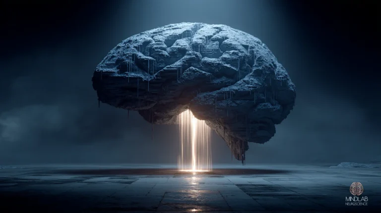
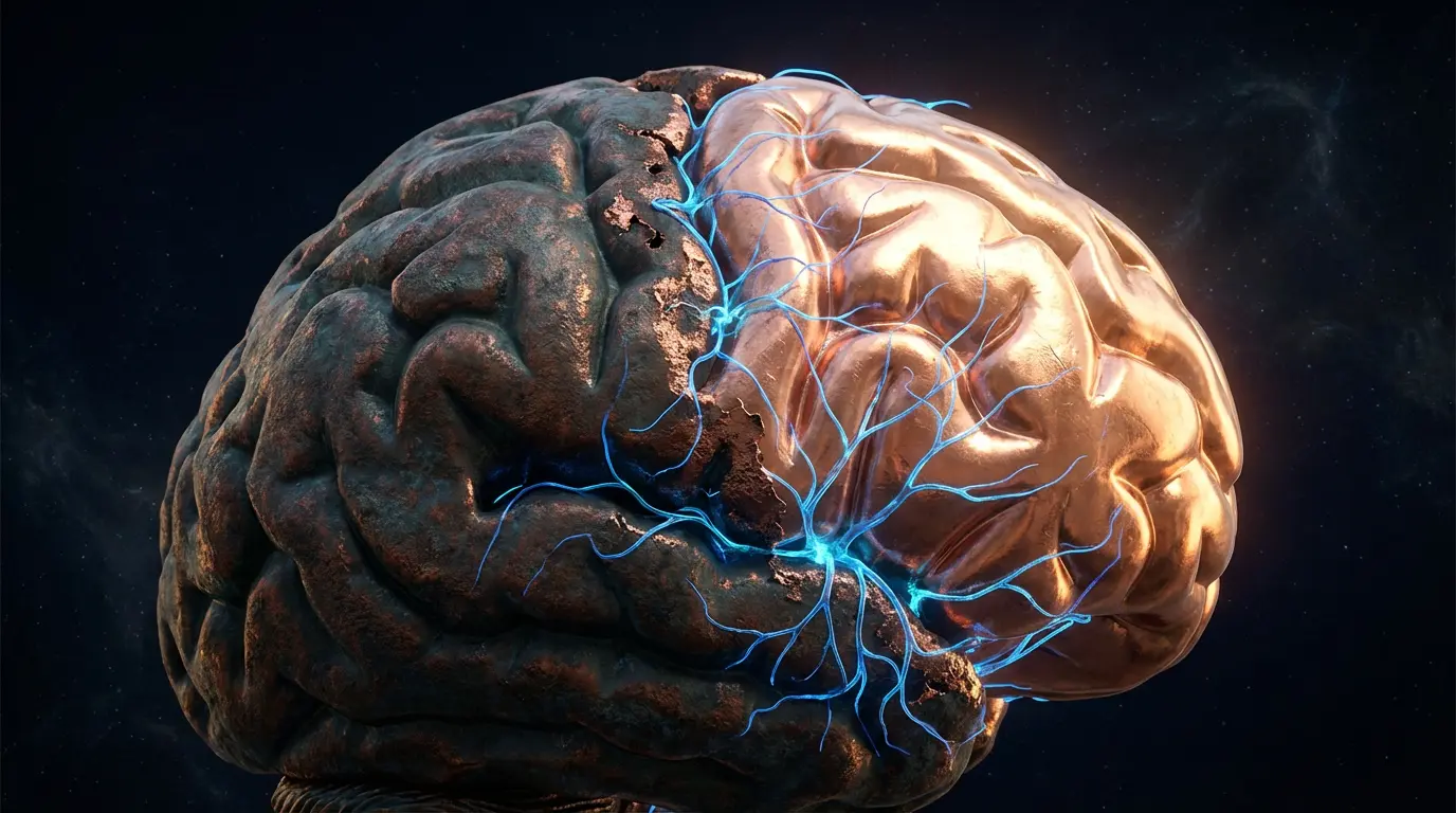
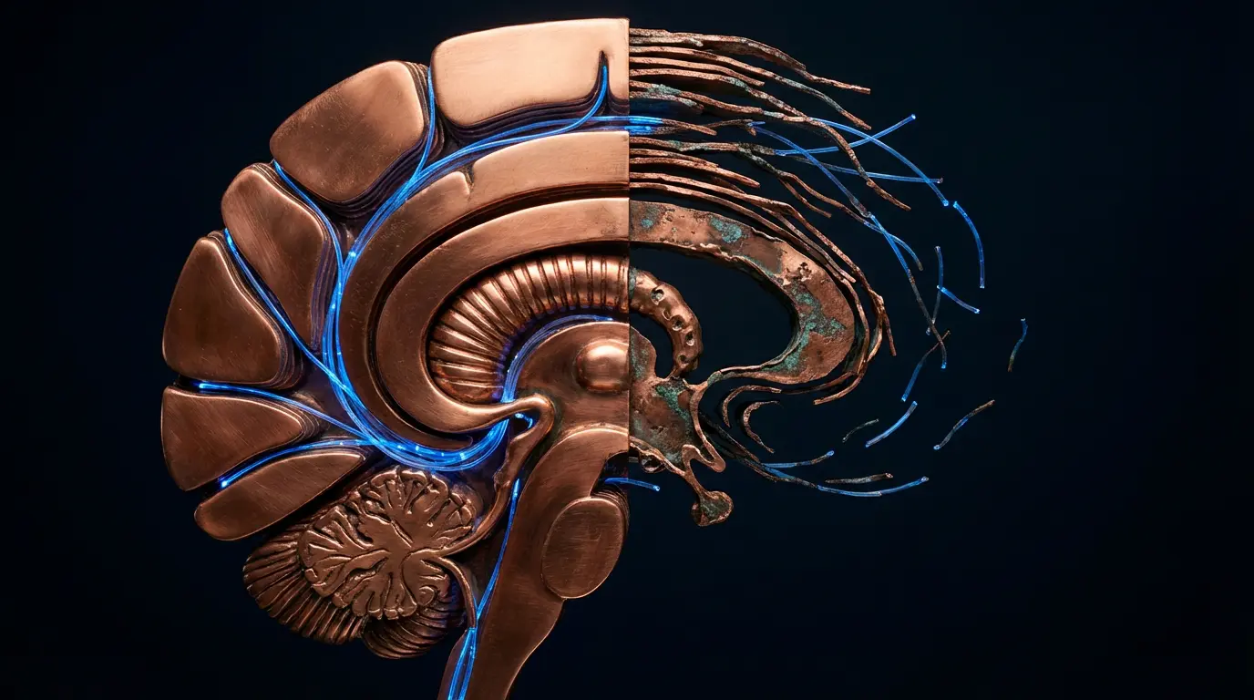
The brain thins quietly under sustained drinking, and the changes surface as behavior. Most people mistake the damage for personality long before they recognize it as something the brain can recover from.
What Structural Damage Does Chronic Alcohol Use Cause?
Chronic alcohol consumption produces measurable structural deterioration in two of the brain’s most functionally significant regions: the prefrontal cortex and the hippocampus. Neuroimaging studies consistently show reduced gray matter volume in both areas among individuals with sustained heavy drinking histories, with the degree of atrophy correlating to duration and quantity of use.

The prefrontal cortex damage manifests as progressive thinning of cortical gray matter, the dense outer layer where complex cognition occurs. Individuals with alcohol use disorder show substantial reductions in prefrontal cortex volume compared with age-matched controls. This structural loss directly impairs the neural circuits governing impulse control, future planning, emotional regulation, and social cognition.
The hippocampal damage follows a parallel trajectory. The hippocampus, essential for converting short-term experiences into long-term memories and for spatial navigation, shows significant volume reduction with chronic alcohol exposure. Imaging studies find that heavy drinkers show significantly smaller hippocampal volumes than non-drinking controls, with the degree of atrophy predicting performance deficits on memory tasks.
This is the moment when someone realizes the sharp mind they relied on feels like it is fading, and they cannot tell whether it is aging, stress, or the drinking. The word that was on the tip of the tongue but never arrived. The project that used to take focus for two hours but now requires four. The mounting suspicion that something has changed, paired with the reluctance to name the most obvious variable.
What the research does not always capture is the behavioral cascade these structural changes produce. When I work with individuals whose drinking history spans years, the presenting complaints are rarely about alcohol itself. They describe difficulty maintaining focus at work, strained relationships they cannot explain, emotional reactions that feel disproportionate, and a persistent sense of cognitive fog. These are not vague complaints, they are the predictable downstream effects of prefrontal and hippocampal atrophy. The neural architecture that would normally buffer stress, contextualize social interactions, and maintain working memory has been physically diminished.
What I find particularly telling in my practice is how often individuals have adapted around these deficits without recognizing them as deficits. They have restructured their entire work life around the cognitive fog (shorter meetings, more delegation, avoiding tasks that require sustained concentration) and attributed the adjustments to “getting older” or “being busy.” When I map these behavioral shifts against the neuroimaging data, the pattern is unmistakable: the compensatory behaviors track precisely with the regions showing the most volume loss.
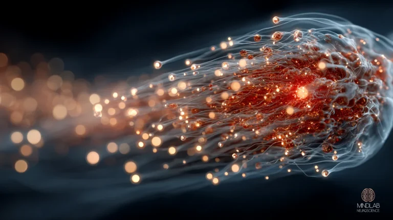
How Does Alcohol Hijack the Brain’s Reward System?
Alcohol triggers dopamine surges in the nucleus accumbens, the brain’s primary reward-processing center, that are significantly larger than those produced by natural rewards like food, social connection, or accomplishment. This pharmacological amplification of dopamine signaling is the neurological foundation of alcohol’s reinforcing properties and the mechanism through which casual use can transition to compulsive use.
The reinforcement loop operates through a process called incentive sensitization. With repeated alcohol exposure, the mesolimbic dopamine pathway becomes increasingly responsive to alcohol-related cues (the sight of a bar, the sound of a cork, the social context of a gathering) while simultaneously becoming less responsive to natural dopamine triggers. This sensitization can develop within weeks of regular consumption and persists for months after cessation.
One pattern I observe repeatedly in my practice, and one that rarely appears in the clinical literature, is what I call the competence trap. High-functioning individuals whose drinking has progressed past moderate use almost never present with the classic signs of alcohol dependence. Instead, they present with a narrowing of identity. Their social life has gradually consolidated around contexts where drinking is present. Their stress management toolkit has contracted to a single instrument. They have not lost control in any visible way, but if you map their dopamine landscape, the terrain has flattened dramatically. The nucleus accumbens is responding to one signal with full intensity and everything else at a fraction. They are still performing, but the neurological margin between performing and unraveling has thinned to a degree they cannot see from the inside.
The downstream consequence is a progressive narrowing of what the brain registers as rewarding. Activities that once generated satisfaction (exercise, creative work, meaningful conversation) produce diminished dopamine responses because the nucleus accumbens has recalibrated its baseline around alcohol’s artificially elevated signal. This is not a failure of motivation or discipline. It is a structural recalibration of the reward circuitry that makes alcohol disproportionately salient relative to every other source of reinforcement in a person’s life.
If you have noticed that the things you used to enjoy (the morning run, the weekend project, the conversation with a friend) feel flat in a way you cannot quite explain, this is the mechanism. It is not depression in the traditional sense. It is a reward system that has been recalibrated to treat a pharmacological signal as the baseline, making everything else feel like it is playing at half volume. The 8pm decision to pour the drink is not really about relaxation, it is the nucleus accumbens recognizing that the one reliable source of a full-strength dopamine signal is within reach.
This recalibration also explains the irritability, restlessness, and anhedonia that characterize early sobriety. The brain’s reward system has been conditioned to expect a dopamine signal of a magnitude that natural experiences cannot produce, and the period of recalibration, while the system gradually restores sensitivity to normal reward levels, is neurologically demanding.
Can the Brain Recover From Alcohol-Related Damage?
The brain retains a remarkable capacity for structural and functional recovery after sustained cessation of alcohol use, though the timeline and extent of recovery depend on the duration and severity of prior exposure. Neuroimaging evidence demonstrates measurable restoration of cortical volume, hippocampal neurogenesis, and normalization of neurotransmitter receptor density within months of abstinence.
Longitudinal imaging across the first year of sustained sobriety shows that prefrontal cortex gray matter volume increases significantly within the first two months, with continued recovery observed through month twelve. Hippocampal volume showed a parallel trajectory, with measurable increases beginning around month three.
The neurogenesis data is particularly significant. The hippocampus is one of the few brain regions where new neurons continue to be generated throughout adulthood, a process called adult neurogenesis. Chronic alcohol exposure suppresses hippocampal neurogenesis, but studies in both animal models and human neuroimaging have demonstrated that this suppression reverses after alcohol cessation. Studies of protracted abstinence document that hippocampal neurogenesis rebounds after alcohol cessation, with structural recovery following over subsequent months.
In my practice, I observe that the individuals who recover most completely are those who pair cessation with active cognitive engagement, deliberate exposure to novel learning, complex problem-solving, and structured social interaction. The brain’s recovery is not purely passive; it responds to demand. Neural circuits that are challenged during the recovery window rebuild more robustly than those that sit dormant. This is the foundational principle behind Real-Time Neuroplasticity™: recovery is not merely the absence of alcohol but the active reconstruction of functional neural architecture, directed at the precise moments when the brain is making its most consequential decisions.
What distinguishes this approach from conventional recovery models is timing and specificity. Traditional interventions occur in clinical settings, removed from the environments where the actual behavioral patterns fire. Through my NeuroConcierge program, I work with individuals inside their real decision environment, the evening transition when the pour becomes automatic, the social setting where the drink is the default stress regulator, the morning-after window when the prefrontal cortex is rebuilding its daily authority. Intervening at these inflection points, rather than reflecting on them after the fact, produces structural change that generalizes across contexts because the neural circuits are being rebuilt under load, not in isolation.
The dopamine system shows its own recovery trajectory. Receptor density in the nucleus accumbens gradually normalizes over three to six months, though full restoration of natural reward sensitivity may take longer depending on the duration of prior use. The practical implication is that the anhedonia and motivational deficits of early sobriety are temporary: the hardware is rebuilding, not permanently damaged.
If you are in the early weeks or months of cessation and the world still feels gray, if music does not land the way it used to, if accomplishments feel hollow, if you are wondering whether the flatness is permanent: it is not. What you are experiencing is the neurological equivalent of recalibration. The brain is restoring its sensitivity to normal-magnitude signals after months or years of artificially amplified ones. The timeline is not instant, but the trajectory is measurable, and the individuals I work with consistently describe a point, often around month three or four, where color begins returning to experience in a way they had forgotten was possible.
References
- Agartz, I., Momenan, R., Rawlings, R.R., Kerich, M.J., & Hommer, D.W. (1999). Hippocampal volume in patients with alcohol dependence. Archives of General Psychiatry, 56(4), 356–363. https://doi.org/10.1001/archpsyc.56.4.356
- Nixon, K., & Crews, F.T. (2004). Temporally specific burst in cell proliferation increases hippocampal neurogenesis in protracted abstinence from alcohol. Journal of Neuroscience, 24(43), 9714–9722. https://pubmed.ncbi.nlm.nih.gov/15509760/
- Volkow, N.D., Wang, G.J., Fowler, J.S., & Tomasi, D. (2012). Addiction circuitry in the human brain. Annual Review of Pharmacology and Toxicology, 52, 321–336. https://doi.org/10.1146/annurev-pharmtox-010611-134625
- Sullivan, E.V., & Pfefferbaum, A. (2005). Neurocircuitry in alcoholism: A substrate of disruption and repair. Psychopharmacology, 180(4), 583–594. https://pubmed.ncbi.nlm.nih.gov/15834536/
- Crews, F.T., & Nixon, K. (2009). Mechanisms of neurodegeneration and regeneration in alcoholism. Alcohol and Alcoholism, 44(2), 115–127. https://doi.org/10.1093/alcalc/agn079
Frequently Asked Questions
How quickly does alcohol begin affecting brain function after consumption?
Alcohol crosses the blood-brain barrier within 60 seconds of entering the bloodstream and begins altering neurotransmitter activity almost immediately. The initial neurological effects, enhanced GABA signaling and suppressed glutamate transmission, produce measurable changes in prefrontal cortex function within five to ten minutes. Blood alcohol concentration peaks approximately 30 to 90 minutes after consumption depending on body composition, food intake, and metabolic rate, but the neurochemical disruption begins well before peak levels are reached.
Is moderate drinking safe for brain health?
Even moderate alcohol consumption produces detectable neurological effects, though the long-term structural implications remain a subject of active research. A 2022 analysis of brain imaging from over 36,000 adults found that any level of alcohol consumption was associated with reduced brain volume, with the relationship becoming steeper at higher intake levels. The neurological effects of alcohol do not appear to have a safe threshold below which no measurable impact occurs, though individual vulnerability varies based on genetics, age, and baseline neural architecture.
How long does it take for the brain to recover after stopping alcohol use?
Structural brain recovery follows a staged timeline after sustained cessation. Prefrontal cortex gray matter volume shows measurable increases within the first two months of abstinence, while hippocampal recovery typically begins around month three. Dopamine receptor density in the nucleus accumbens normalizes over three to six months. Full functional recovery, including restoration of natural reward sensitivity and executive function, may take 12 to 18 months depending on prior duration and severity of use, though significant improvement is observable much earlier in the process.
Can neuroplasticity-based intervention accelerate recovery from alcohol’s neurological effects?
Neuroplasticity-based intervention can meaningfully accelerate the brain’s structural recovery by providing targeted cognitive demands that drive circuit reconstruction during the recovery window. The brain rebuilds neural pathways in response to specific demands placed upon them. Deliberate exposure to complex decision-making, emotional regulation challenges, and novel learning experiences stimulates the prefrontal and hippocampal circuits that alcohol damaged. Dr. Ceruto’s Real-Time Neuroplasticity™ methodology leverages this demand-driven plasticity by intervening during real-time behavioral moments, not in a clinical office after the fact, but embedded in the individual’s actual decision environment when the brain is most receptive to lasting architectural change.
Does alcohol affect everyone’s brain the same way?
Alcohol’s neurological effects vary significantly across individuals due to genetic polymorphisms in alcohol-metabolizing enzymes, baseline neurotransmitter receptor density, age of first exposure, and pre-existing neural architecture. Individuals with genetic variants that produce slower alcohol metabolism experience prolonged neurotoxic exposure per drinking episode. Additionally, the adolescent and young adult brain, where prefrontal cortex myelination continues until approximately age 25, is disproportionately vulnerable to alcohol-related structural damage compared to the fully mature adult brain.
What the First Conversation Looks Like
If you are reading this because you have started to wonder whether the nightly drink is doing more than relaxing you, if the fog is getting harder to explain away, if the sharpness you used to count on feels like it is slipping, if the 8pm pour has shifted from a choice to something closer to a default, that recognition is not anxiety. It is accuracy. Your brain is telling you what the research confirms.
A strategy call with Dr. Ceruto is a private, one-hour neurological assessment, not a clinical intake, not a sobriety lecture, not a motivational conversation. Dr. Ceruto evaluates the specific circuits driving your current pattern: the reward system recalibration, the prefrontal thinning, the stress-response dysregulation, the compensatory behaviors you may have built around deficits you have not yet named. She maps the architecture and tells you directly what is structurally reversible, what the recovery timeline looks like, and whether her Real-Time Neuroplasticity™ methodology is the right intervention for where your brain is right now.
Most people who schedule this call have already tried willpower, moderation rules, and self-monitoring. What they have not had is someone who can show them exactly which neural circuits are driving the pattern and what it takes to rebuild them. That is what this conversation provides.
Crisis & mental-health support resources
If you are struggling with alcohol use, free and confidential support is available right now.
- SAMHSA National Helpline: call 1-800-662-HELP (4357) for 24/7 free, confidential support and referrals.
- 988 Suicide & Crisis Lifeline: call or text 988 if you are in emotional crisis.
- If you are in immediate danger, call 911 or go to your nearest emergency room.