Key Takeaways
- Chronic low-grade neuroinflammation differs from the brain’s acute immune response, silently eroding cognition over years.
- Persistent pro-inflammatory cytokines (IL-6, TNF-alpha, IL-1-beta) disrupt synaptic plasticity, suppress hippocampal neurogenesis, impair prefrontal function.
- Microglial cells “primed” by repeated inflammatory insults produce exaggerated responses to even minor subsequent triggers.
- Blood-brain barrier degrades under inflammatory pressure, letting peripheral immune signals amplify central nervous system inflammation.
- Structured exercise, restorative sleep, and anti-inflammatory nutrition measurably reduce neuroinflammatory markers and support cognitive resilience.
Every second of every day, billions of immune cells patrol the brain, maintaining the delicate equilibrium that sustains thought, memory, and focus. That equilibrium is what protects the neural architecture that sustains cognition. When that surveillance system shifts from protective vigilance to sustained aggression, the consequences ripple across every cognitive domain: from the sharpness of a recalled name to the clarity of a complex decision.
Acute Protection Versus Chronic Destruction
The brain’s inflammatory response evolved as an exquisitely calibrated defense mechanism. Acute inflammation following injury or infection mobilizes immune resources rapidly, clears damaged tissue, and initiates repair processes that restore neural function within days. This protective cascade is precisely time-limited: molecular braking signals shut it down once the threat has passed.
Chronic low-grade neuroinflammation operates under entirely different rules. Rather than a sharp spike followed by resolution, the immune machinery remains partially activated for weeks, months, or years at a level too low to produce obvious signs yet high enough to cause cumulative structural damage. This sustained smoldering state does not resolve on its own because the signals that should terminate the response become blunted by persistent activation. The result is a brain environment where the very mechanisms designed to protect neurons gradually turn against them.
Understanding this distinction matters because the interventions that support acute immune function (rest, adequate nutrition, temporary reduction of demands) differ substantially from the sustained lifestyle architecture required to counteract chronic neuroinflammatory processes. Whereas acute inflammation follows a predictable arc with identifiable resolution markers, chronic neuroinflammation operates without clear temporal boundaries, often requiring objective biomarker assessment to detect and quantify.
| Dimension | Acute inflammation | Chronic low-grade neuroinflammation |
|---|---|---|
| Time course | Sharp spike, then resolution within days | Smolders for weeks, months, or years |
| Resolution signals | Molecular brakes shut it down once the threat passes | Termination signals blunted by persistent activation |
| Microglial state | Activate, clear debris, return to surveillance | “Primed”: exaggerated response to minor triggers |
| Effect on neurons | Clears damage and initiates repair | Disrupts plasticity, suppresses neurogenesis, erodes prefrontal function |
| Net role | Protective and essential | Cumulative structural damage beneath awareness |
Microglia: When Sentinels Become Aggressors
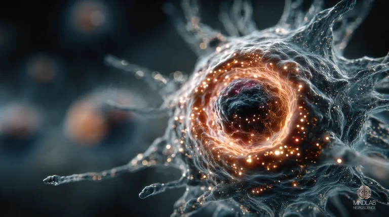
Microglia constitute roughly ten percent of all brain cells and serve as the central nervous system’s primary immune surveillance network. Under healthy conditions, these cells continuously extend and retract fine processes, monitoring synaptic activity and clearing cellular debris with remarkable precision. They also participate actively in synaptic pruning during development and in maintaining the synaptic connections that underlie learning throughout adulthood.
When microglia detect danger signals (from misfolded proteins, cellular damage, or peripheral immune messengers) they shift into an activated state, releasing pro-inflammatory cytokines and reactive oxygen species intended to neutralize threats. In the acute context, this response is both appropriate and essential. The problem emerges when activation becomes chronic. Repeated or sustained inflammatory triggers produce a state researchers term “microglial priming,” where these cells adopt a heightened baseline reactivity. Primed microglia respond to even minor secondary insults with disproportionate inflammatory output, creating a positive feedback loop that amplifies neural damage far beyond what the original trigger warranted.
This priming phenomenon helps explain why individuals with existing low-grade inflammation (whether from metabolic conditions, chronic stress, or sedentary lifestyles) experience more severe cognitive consequences from subsequent inflammatory events such as infections or surgical procedures.
The Cytokine Cascade and Synaptic Disruption
Three pro-inflammatory cytokines dominate the neuroinflammatory landscape: interleukin-6 (IL-6), tumor necrosis factor-alpha (TNF-alpha), and interleukin-1-beta (IL-1-beta). Each contributes distinct mechanisms of cognitive disruption when chronically elevated.
IL-6, produced by activated microglia and astrocytes, crosses the blood-brain barrier bidirectionally and activates the JAK/STAT signaling pathway. At acutely elevated levels, IL-6 can paradoxically support neuroprotection. However, chronically elevated IL-6 shifts this signaling toward trans-signaling pathways that promote sustained inflammation and have been directly associated with accelerated cognitive decline, particularly in memory domains.
TNF-alpha exerts its effects partly through the modulation of glutamate receptors at the synapse. Sustained TNF-alpha elevation increases excitatory neurotransmission while simultaneously reducing inhibitory signaling, creating an excitatory-inhibitory imbalance that impairs the precise synaptic calibration required for long-term potentiation and memory consolidation. This imbalance also increases vulnerability to excitotoxicity, where excessive glutamate signaling damages or destroys neurons.
IL-1-beta directly suppresses hippocampal neurogenesis: the brain’s capacity to generate new neurons in the region most critical for memory formation. Research has demonstrated that sustained IL-1-beta exposure reduces the production of new neurons in the hippocampal dentate gyrus and shifts neural precursor cell differentiation away from neuronal lineages toward glial lineages. This means the brain not only produces fewer new neurons under chronic inflammatory conditions but also redirects its limited regenerative capacity toward non-neuronal cell types.
Blood-Brain Barrier Compromise
The blood-brain barrier represents one of the body’s most sophisticated biological filters, composed of tightly joined endothelial cells that selectively regulate which molecules enter the brain from peripheral circulation. Under normal conditions, this barrier prevents the vast majority of circulating immune cells and inflammatory molecules from reaching neural tissue.
Chronic inflammation degrades this barrier through multiple convergent mechanisms. TNF-alpha and IL-1-beta weaken tight junction proteins between endothelial cells, creating gaps that permit peripheral inflammatory molecules to enter the brain parenchyma. Simultaneously, these cytokines upregulate adhesion molecules on the barrier’s surface, actively recruiting circulating immune cells, neutrophils and T-cells, to cross into the central nervous system.
Once peripheral immune cells breach the barrier, they release additional pro-inflammatory cytokines and chemokines within the brain itself, establishing a self-amplifying cycle. Peripheral inflammation generates central inflammation, which further weakens the barrier, which permits more peripheral inflammation to enter. This vicious cycle helps explain why systemic inflammatory conditions (metabolic dysfunction, chronic stress, poor sleep) produce cognitive effects that seem disproportionate to their apparent severity. The brain is not merely registering distant inflammation; it is being directly invaded by it.
Prefrontal Function and Executive Erosion
While hippocampal damage from neuroinflammation receives significant research attention, the effects on the prefrontal cortex are equally consequential for everyday cognitive performance. The prefrontal cortex governs executive functions: working memory, attentional control, planning, decision-making, and the ability to regulate emotional responses.
Chronic neuroinflammation disrupts prefrontal function through several mechanisms. Activated microglia and elevated cytokines within prefrontal regions alter the excitatory-inhibitory balance that underlies precise neural computation. Research has demonstrated that inflammatory processes in the prefrontal cortex shift processing resources away from deliberate, reflective evaluation and toward faster, more reactive response patterns. This manifests as difficulty sustaining attention, impaired working memory, reduced cognitive flexibility, and compromised executive function under demanding conditions.
The prefrontal cortex is particularly vulnerable to inflammatory disruption because the dendritic arbors of its pyramidal neurons, the elaborate branching structures that receive synaptic inputs, are among the most complex in the brain. This complexity enables sophisticated information integration but also creates a larger surface area susceptible to inflammatory damage. Chronic cytokine exposure causes measurable dendritic retraction and spine loss in prefrontal neurons, physically reducing the brain’s capacity for the kinds of nuanced processing that distinguish expert-level thinking from reflexive responding. Over time, this structural degradation shifts the balance of cognitive control toward subcortical circuits, producing a measurable decline in the deliberate, goal-directed reasoning that prefrontal networks are uniquely evolved to support.
Acute inflammation is the brain defending itself; chronic inflammation is that defense left running until it consumes what it was built to protect.
Exercise as Neuroinflammatory Medicine
Physical exercise produces what may be the most robust anti-neuroinflammatory effect documented in lifestyle research. The mechanisms are both direct and indirect, operating through multiple complementary pathways.
During acute exercise, skeletal muscles release IL-6 in a context-dependent manner that paradoxically reduces chronic inflammation. Exercise-derived IL-6 activates anti-inflammatory downstream cascades, stimulating the release of interleukin-1 receptor antagonist (IL-1RA) and interleukin-10 (IL-10), both of which suppress pro-inflammatory signaling. Over time, regular exercise training reduces baseline levels of circulating TNF-alpha, IL-1-beta, and chronic IL-6, fundamentally recalibrating the inflammatory set point.
Within the brain, exercise directly modulates microglial phenotype, shifting activated microglia from pro-inflammatory states toward neuroprotective configurations that support synaptic maintenance and debris clearance without excessive cytokine release. Exercise also upregulates brain-derived neurotrophic factor (BDNF), which promotes hippocampal neurogenesis and counteracts the suppressive effects of chronic inflammation on new neuron production. The combined effect is a measurable preservation of both hippocampal and prefrontal structure and function, even in populations with elevated baseline inflammatory risk.
Sleep Architecture and Inflammatory Regulation
Sleep serves as the brain’s primary anti-inflammatory maintenance window. During deep slow-wave sleep, the glymphatic system, a recently characterized waste-clearance network, flushes inflammatory byproducts, misfolded proteins, and cellular debris from the interstitial spaces between neurons. Disrupted or insufficient sleep directly impairs this clearance process while simultaneously activating pro-inflammatory pathways.
Research has established that even modest sleep deprivation produces measurable increases in circulating TNF-alpha, IL-6, and IL-1-beta, and activates microglial cells in ways that mirror chronic neuroinflammatory states. Sleep loss also activates the NF-kB signaling pathway, a master regulator of inflammatory gene expression, in ways that persist even after recovery sleep, suggesting a cumulative inflammatory debt that builds with repeated sleep disruption.
The relationship is bidirectional: neuroinflammation itself disrupts sleep architecture by altering the neurotransmitter balances that regulate sleep-wake transitions, creating another self-reinforcing cycle. Breaking this cycle requires not merely increasing sleep duration but restoring the structural quality of sleep, particularly the deep slow-wave stages during which glymphatic clearance is most active.
Nutritional Modulation of Inflammatory Tone
Dietary patterns exert profound and sustained influence over systemic and central inflammatory status. The Mediterranean dietary pattern (characterized by high consumption of omega-3 fatty acids from fish, polyphenol-rich extra-virgin olive oil, diverse vegetables, and limited processed foods) has been repeatedly associated with reduced circulating inflammatory markers and preserved cognitive function across aging populations.
The mechanisms operate at multiple levels. Omega-3 fatty acids, particularly docosahexaenoic acid (DHA) and eicosapentaenoic acid (EPA), are incorporated into neuronal membranes where they serve as precursors for specialized pro-resolving mediators: molecules that actively terminate inflammatory responses rather than merely suppressing them. Polyphenols from olive oil, berries, and leafy greens inhibit NF-kB activation and reduce microglial pro-inflammatory cytokine production. Dietary fiber supports gut microbial communities that produce short-chain fatty acids with direct anti-neuroinflammatory properties, operating through the gut-brain axis.
Conversely, ultra-processed food patterns high in refined sugars and industrial seed oils promote systemic inflammation through multiple pathways, including endotoxemia, the leakage of bacterial products from the gut into circulation, which directly triggers microglial activation via toll-like receptors. The cumulative evidence indicates that dietary choices represent not an adjunct to brain health and optimization but a foundational determinant of the brain’s inflammatory environment.
About the Author
Founder & CEO of MindLAB Neuroscience, Dr. Sydney Ceruto is the pioneer of Real-Time Neuroplasticity™: a proprietary methodology that durably rewires the neural pathways driving behavior, decisions, and emotional responses.
Dr. Ceruto holds a PhD in Behavioral & Cognitive Neuroscience (NYU) and Master’s degrees in Clinical Psychology and Business Psychology (Yale University). Lecturer, Wharton Executive Development Program, University of Pennsylvania.
Chronic neuroinflammation does not announce itself with dramatic episodes: it operates beneath conscious awareness, gradually eroding the neural infrastructure that supports clear thinking, reliable memory, and sound decision-making. The neuroscience is clear: the brain’s inflammatory environment is not fixed but responsive to deliberate, sustained lifestyle choices. If persistent cognitive fog, difficulty concentrating, or a sense that your mental sharpness has diminished has become your baseline, these may reflect neuroinflammatory processes that targeted neural intervention can address. Book a Strategy Call to learn how MindLAB Neuroscience applies Real-Time Neuroplasticity™ to restore cognitive performance at the level where it originates: the brain itself.
What is chronic neuroinflammation?
It is a low-grade, sustained activation of the brain’s immune system, distinct from the acute, time-limited inflammation that follows injury or infection. Instead of spiking and resolving, it smolders for months or years at a level too low to notice yet high enough to cause cumulative damage, because the signals that should shut it down become blunted.
How does inflammation impair memory and focus?
Three pro-inflammatory cytokines, IL-6, TNF-alpha, and IL-1-beta, do most of the damage. TNF-alpha unbalances excitatory and inhibitory signaling, IL-1-beta suppresses hippocampal neurogenesis, and chronically elevated IL-6 tracks with memory decline. Together they disrupt synaptic plasticity and degrade prefrontal control of attention, working memory, and decision-making.
What is microglial priming?
Microglia are the brain’s resident immune cells. Repeated or sustained inflammatory triggers can leave them “primed”: set at a heightened baseline reactivity. Primed microglia then overreact to even minor later insults, releasing disproportionate inflammatory output. That is why people with existing low-grade inflammation often suffer worse cognitive effects from a subsequent infection or surgery.
Can chronic brain inflammation be reduced?
Yes, and the levers are largely behavioral. Regular exercise lowers baseline TNF-alpha, IL-1-beta, and IL-6 and shifts microglia toward neuroprotective states. Restorative slow-wave sleep drives glymphatic clearance of inflammatory debris. A Mediterranean-style diet rich in omega-3s and polyphenols supplies pro-resolving mediators and inhibits inflammatory signaling: together measurably reducing neuroinflammatory markers.
How are sleep and inflammation connected?
Bidirectionally. Even modest sleep loss raises circulating TNF-alpha, IL-6, and IL-1-beta and activates microglia, while deep slow-wave sleep is when the glymphatic system flushes inflammatory byproducts. Inflammation in turn disrupts the neurotransmitter balance that regulates sleep: a self-reinforcing cycle broken by restoring slow-wave sleep quality, not just duration.
References
- Heneka, M. and others. Neuroinflammation in Alzheimer’s disease. The Lancet Neurology, 14(4), 388-405, 2015. https://doi.org/10.1016/s1474-4422(15)70016-5
- Salter, M. and Stevens, B. Microglia emerge as central players in brain disease. Nature Medicine, 23(9), 1018-1027, 2017. https://doi.org/10.1038/nm.4397
- Perry, V. and Holmes, C. Microglial priming in neurodegenerative disease. Nature Reviews Neurology, 10, 217-224, 2014. https://doi.org/10.1038/nrneurol.2014.38
- Heppner, F., Ransohoff, R., and Becher, B. Immune attack: the role of inflammation in Alzheimer disease. Nature Reviews Neuroscience, 16(6), 358-372, 2015. https://doi.org/10.1038/nrn3880
- Ransohoff, R. How neuroinflammation contributes to neurodegeneration. Science, 353(6301), 777-783, 2016. https://doi.org/10.1126/science.aag2590
- Monje, M., Toda, H., and Palmer, T. Inflammatory blockade restores adult hippocampal neurogenesis. Science, 302(5651), 1760-1765, 2003. https://doi.org/10.1126/science.1088417